Figure 6.
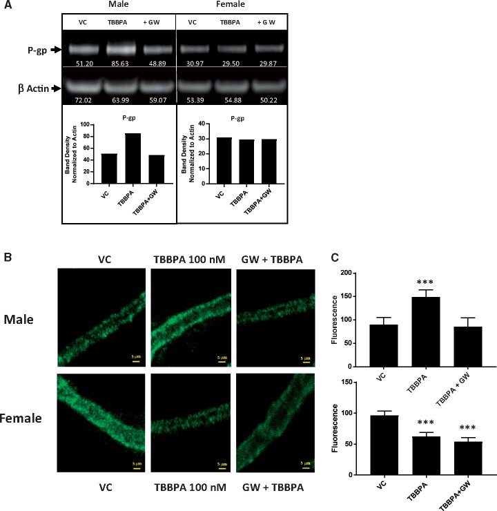
Inhibition of PPAR-γ blocks TBBPA mediated increases in P-gp expression in males. A, Western blot containing proteins from male (left) and female (right) rat brain capillaries. Non-treated vehicle controls (NT), TBBPA 100 nM exposed capillaries (TBBPA), and co-treated with TBBPA + 10 nM GW 9668 (+ GW). B, (top) Immunofluorescent images showing TBBPA increases are blocked by inhibition of PPAR-γ in males. Band density was determined by densitometry using ImageJ software. White numbers under P-gp bands in blot were normalized to the density of actin bands to control for minor loading variances. Graphs depict normalized density values of P-gp band in Western blot. B, (bottom) Immunofluorescent images showing TBBPA decreases in females are insensitive to PPAR-γ inhibition. Graphical analysis of P-gp immunofluorescence in males (C, top) and females (C, bottom). Shown are mean fluorescence ± SEM for 10–20 capillaries from single preparation (pooled brains from 3 to 5 rats, 15–20 weeks old). ***p < .001, significantly different than control. Scale bar equals 10 µm in length. Abbreviations: P-gp, P-glycoprotein; TBBPA, tetrabromobisphenol A.

