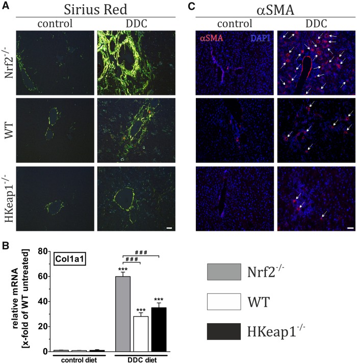
Figure 3.
Nrf2 prevents liver fibrosis and stellate cell activation. Nrf2−/−, WT, and HKeap1−/− were fed for 4 weeks with normal chow (control) or 0.1% DDC-containing diet (DDC). Afterward, liver tissue was collected and prepared as described in the Materials and Methods section. (A) Sirius-red staining to visualize fibrotic changes. (B) mRNA expression level of Col1a1 as a fibrotic marker using qRT-PCR. (C) Immunofluorescent staining against αSMA for the detection of activated stellate cells. A and C show representative pictures out of n = 6; data = mean + SEM; n = 6; ***p < .001 versus control diet; ###p < .001 as indicated.

