Figure 6.
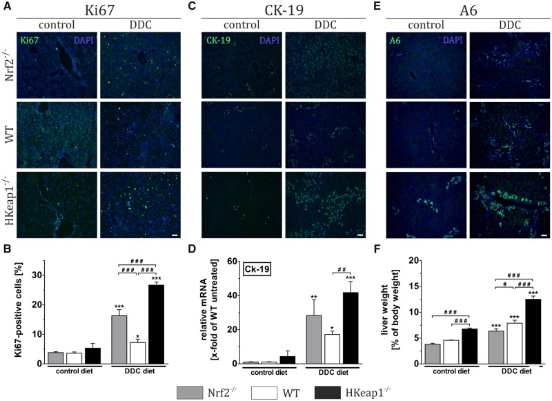
Nrf2 enhances liver regeneration due to increased oval cell activation. Examination of liver regeneration was conducted by Ki67 staining (A) and quantification of Ki67-positive cells (B). Cholangiocytes/cholangitis were visualized by immunofluorescent labeling of CK-19 protein (C) and further quantified by mRNA expression analysis of Ck-19 (D). Examination of oval cell activation was conducted by immunofluorescent detection using an antibody against A6 (E). Relative liver weights are depicted in (F) as calculated, n ≥ 8. Representative images out of n = 6 are shown (scale = 100 µm). Data = mean + SEM, *p < .05, **p < .01, and ***p < .001 versus control diet; ###p < .001 as indicated.

