Figure 2.

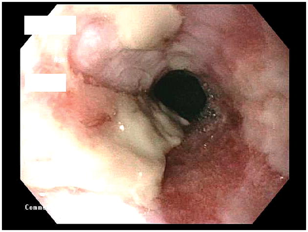
Endoscopic photo of the distal esophagus showing LA-D esophagitis. The extensive mucosal breaks involve most (>75%) of the circumference of the distal esophagus. Much of the broken mucosal surface is covered by dense, whitish exudate.

Endoscopic photo of the distal esophagus showing LA-D esophagitis. The extensive mucosal breaks involve most (>75%) of the circumference of the distal esophagus. Much of the broken mucosal surface is covered by dense, whitish exudate.