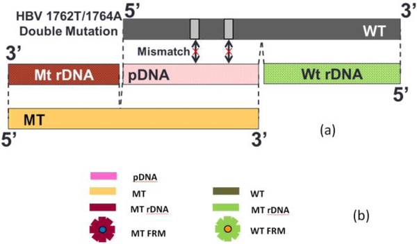
Fig. 2.
(a) A schematic of the relationship between probe DNA (pDNA), mutant (MT) target DNA (tDNA), wild type (WT), MT reporter DNA (MT rDNA), and WT rDNA (WT rDMA) for HBV 1762/1764 double mutation and (b) a schematic representation of (a) pDNA, MT, MT rDNA, and blue MT fluorescent reporter microsphere (MT FRM), and WT, WT rDNA, and orange WT fluorescent reporter microsphere (WT FRM).

