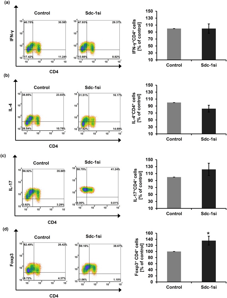
Fig 3. Flow cytometric analysis of normal T helper subsets upon direct co-culture with Sdc-1-silenced SUM-149 cells.
PBMCs isolated from normal subjects were co-cultured with Sdc-1-silenced SUM-149 cells for 48 hours. Lymphocytes were then stained with labeled antibodies against CD4-FITC, IFN-γ-PE, IL-4-PEcy7, IL-17-PE, and Foxp3-PEcy7. Relative to control cells, tumor Sdc-1 silencing significantly increased the percentage of (d) Treg (Foxp3+CD4+) subsets while did not change the percentage of the other CD4+ T cell subsets (a-c). Left panels of (a-d) are representative flow cytometric analyses of CD4+ T cell subsets. Data shown is representative for a single experiment. Right panels of (a-d) show the quantification of CD4+ T cell subsets as analyzed by flow cytometry. Data represent mean ± SEM, n = 5, * P ≤ 0.05 as determined by Student’s t test.

