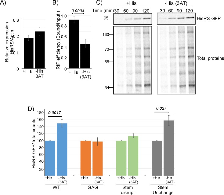
Fig 6. Anticodon mimic–dependent regulation of mRNA binding and HisRS protein synthesis.
A–B) Cells were grown either in the presence of histidine (+His) or without histidine and with 100-mM 3AT (−His) for 1 hour and subjected to GFP-Trap mRNA isolation. A) RT-qPCR analysis of steady-state (Input) HisRS mRNA levels, normalized to Actin levels. B) RIP efficiency (ratio of Bound to Input), as measured by RT-qPCR. Error bars are SEM from two independent biological repeats, and P value was calculated by the dependent samples one-sided t test. See S6 Table for raw data. C) Cells were subjected to 3AT treatment as in A and concomitantly pulsed with 35S-met for the indicated times. Total proteins' sample was set aside, and labeled HisRS was isolated from the remainder by IP. Autoradiographs present the signals of HisRS and Total proteins samples. D) Cells expressing WT or anticodon mimic variants were subjected to a 90-min pulse concomitant with His depletion. Labeled HisRS-GFP was isolated by immunoprecipitation as in C. Histograms present the quantification of the HisRS signals, normalized to the Total proteins signal. Results are from four (for the WT variant) and two (for the anticodon mimic variants) independent biological repeats. Error bars are SEM and P value was calculated by the dependent samples one-sided t test. See S6 Table for raw data. 3AT, 3 aminotriazole; GFP, green fluorescent protein; HisRS, histidyl-tRNA synthetase; IP, immunoprecipitation; RIP, RNA immunoprecipitation; RT-qPCR, reverse transcription quantitative PCR; WT, wild-type.

