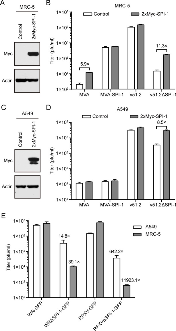
Fig 5. Replication of recombinant MVA, VACV WR, and RPXV in parental or SPI-1-expressing MRC-5 and A549 cells.
(A, C) Expression of SPI-1. MRC-5 (A) and A549 (C) cells were infected with retroviruses that express 2xMyc-SPI-1 or control retroviruses and selected by antibiotic resistance. Western blots with antibody to the Myc tag are shown. The minor Myc band in panel C is assumed to be due to degradation. (B, D) Replication of MVA, MVA-SPI-1, v51.2, v51.2ΔSPI-1 at MOI of 0.01 in control and SPI-1 expressing MRC-5 (B) and A549 (D) cells. (E) A549 and MRC-5 cells were infected at a MOI of 0.01 in triplicate with VACV WR expressing GFP without (WR-GFP) or with a deletion of SPI-1 (WRΔSPI-1-GFP) and RPXV expressing GFP without (RPXV-GFP) or with a deletion of SPI-1 (RPXVΔSPI-1-GFP). In panels B, D and E the cells were infected in triplicate for 48 h and titers were determined in duplicate on CEF. Error bars represent SEM and fold difference in titers are indicated.

