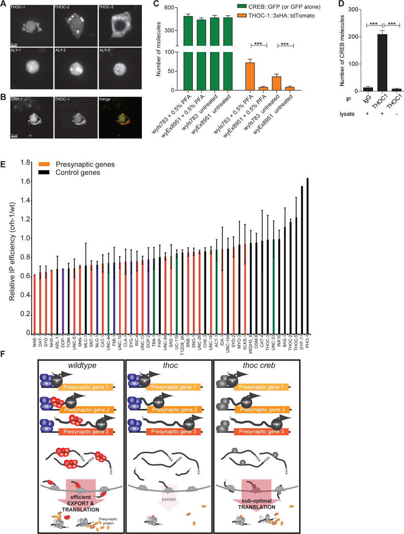
Figure 7: CREB physically interacts with THOC to recruit it onto activity-dependent transcripts.

(A) Localization of GFP-tagged THOC components in PDE cell body.
(B) Co-expression analysis of fluorescently tagged THOC-1::tdTomato and CREB::GFP in PDE neurons.
(C) Quantification of simPULL assay showing that THOC-1 and CREB transiently interact in C. elegans. GFP serves as negative control. ****, p<0.0001 student t-test.
(D) Quantification of simPULL assay showing that THOC1 and CREB transiently interact in mouse striatal brain lysate. No lysate and non-specific IgG antibodies served as negative control. ***, p<0.0001 one-way Anova
(E) Immunoprecipitation of THOC-1-bound RNAs from wt and crh-1 mutant neurons. Red: synaptic genes, purple: postsynaptic genes, blue: cell adhesion molecules, green: cytoskeletal genes, black: other pan-neuronal and control genes.
(F) Current model of how THOC functions to regulate presynaptic gene expression in DA neurons. In wt, CREB recruits THOC onto presynaptic transcripts to facilitate their efficient nuclear export for concerted translation. In THOC mutant background, these transcripts are retained in the nucleus, hence leading to decreased presynaptic proteins and defective synapse assembly. This defect can be alleviated by removal of CREB, thereby allowing for a constitutive backup export pathway to kick in and to re-establish efficient export of presynaptic transcripts.
Averages and SEM are plotted in all graphs.
See also Figure S7.
