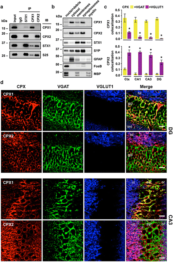
Fig. 1.
Characterization and localization of complexins I (CPX1) and II (CPX2) in human and rat brain tissues. a Immunoprecipitation (IP) with specific antibodies against mouse IgG (as negative control), syntaxin-1 (STX1), complexin-I and complexin-II, using as input human crude cortical (inferior temporal) homogenates from subjects without cognitive impairment, psychiatric or neuropathological conditions. IP products, along with input samples, were resolved by immunoblotting (IB) with specific antibodies against complexins I and II, syntaxin-1, and SNAP-25 (S25). Note that complexin-I/II-IP products were loaded with some lanes of separation from the rest of the samples within the same SDS-gels. Resulting immunoblot captions were edited to cutoff non-informative lanes without altering image levels. b Sequential extraction of nuclear (with cell debris), soluble cytosolic, whole membranal, purified synaptosomal, and myelin-associated proteins from human cortical homogenates. Samples from each fraction were resolved by IB with antibodies against complexins I and II, syntaxin-1, and other markers for synaptic vesicles (synaptophysin [SYP]), cytosol (glial fibrillary acidic protein [GFAP]), nuclei (FosB), and myelin fragments (myelin basic protein [MBP]). Note that MBP strongly labels the nuclear fraction, as heavy myelin fragments precipitate along with cell debris. a–b Masses (in kDa) of prestained markers are indicated on the left side of immunoblots. c Quantitative colocalization analyses between complexin-I/II and each of the vesicular transporters for GABA (VGAT) and glutamate (VGLUT1) using laser-scan confocal microscopy in rat cortex (Ctx) and three hippocampal subfields. Bars represent mean±standard error of n = 4 rats, with at least 24 images analyzed per rat and brain area. *P < 0.001 (paired t-test). d Representative single- or merged-channel confocal images from triple co-immunolabeled rat brain sections described in (c) framing hippocampal dentate gyrus (DG; top panels) and CA3 subfield (bottom panels). Colors were arbitrarily assigned to maximize overlap visualization. Abbreviations: gcl, granule cell layer; hil, hilus; or, stratum oriens; iml, inner molecular layer; pyr, pyralmidal cell layer; r/l, stratum radiatum/lucidum. Scale bars: 10 μm

