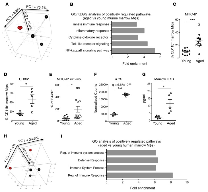
Figure 5. Aged marrow macrophages have a proinflammatory phenotype and elevated IL-1B expression.
(A) PCA plot of young (red) and aged (black) marrow Mφs isolated as shown in Supplemental Figure 3 (n = 3 mice per group). Variance of each PCA is shown on the axes. (B) Upregulated GO and KEGG categories in aged Mφs compared with young (n = 3 mice per group). See also Supplemental Tables 3 and 4 for details. (C and D) Expression of MHC-II (C) and CD86 (D) on Mφs from young and aged mice (n = 5–10 mice per group). (E) Expression of MHC-II in Mφs from BMME in vitro cultures (n = 11–14 wells per group). (F) Expression of Il1b in young and aged Mφs (n = 3 mice per group). (G) Quantification by ELISA of IL-1B protein in marrow of young and aged mice (n = 5 per group). (H) PCA plot of young (red, n = 2) and aged (black, n = 3) human marrow Mφs isolated as shown in Supplemental Figure 4. Variance of each PCA is shown on the axes. (I) Upregulated GO processes in aged human Mφs compared with young (n = 2–3 human marrow per group). Each symbol represents an individual mouse; data represent mean ± SEM. P values, 2-tailed Student’s t test: *P < 0.05, ***P < 0.001.

