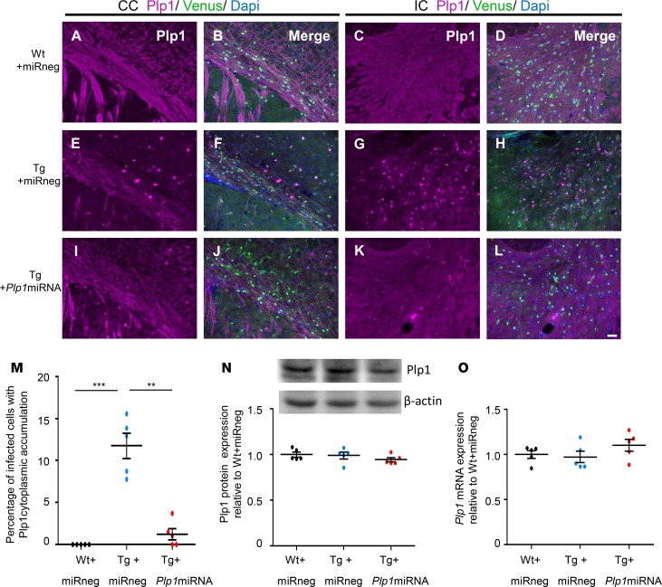
Figure 5. scAAV.CNP.Venus.Plp1miRNA treatment alleviated cytoplasmic accumulation of Plp1 protein in the oligodendrocytes of Plp1-Tg mice.
Coronal sections of P25 mouse brains with Plp1 immunostaining in the CC (A, B, E, F, I, and J) and IC (C, D, G, H, K, and L). The Plp1 expression in Venus-positive oligodendrocytes was detected in Wt+miRneg mice (A–D), Tg+miRneg mice (E–H), and Tg+Plp1miRNA mice (I–L). Scale bar: 50 μm. (M) Proportion of the cells showing Plp1 cytoplasmic accumulation in total Venus-positive cells in the CC and IC (n = 5 mice per group). (N) Representative Western blot analysis of Plp1 expression in scAAV-injected brain tissue of WT and Plp1-Tg mice. β-Actin was used as an internal control. Quantification of the intensity of the Plp1 bands is shown in the graph below (n = 5 mice per group). (O) Quantification of the relative expression of Plp1 mRNA in scAAV-injected brain tissue of WT and Plp1-Tg mice (n = 5 mice per group). Statistical significance was determined using 1-way ANOVA with Bonferroni’s post hoc test. **P < 0.01; ***P < 0.001.

