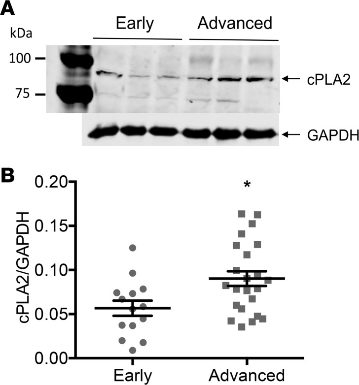
Figure 2. cPLA2 expression was increased in the arteries of advanced- compared with early-stage CVD patients.
(A) Representative cPLA2 and GAPDH Western blots in iliac arteries from early- (n = 3) versus advanced-stage (n = 3) CVD patients. (B) Scatter dot plots (arithmetic mean ± SEM) of cPLA2 protein expression normalized to GAPDH in iliac arteries from early- (n = 14) versus advanced-stage (n = 23) CVD patients. *P < 0.05 vs. early-stage CVD group (unpaired Mann-Whitney test).

