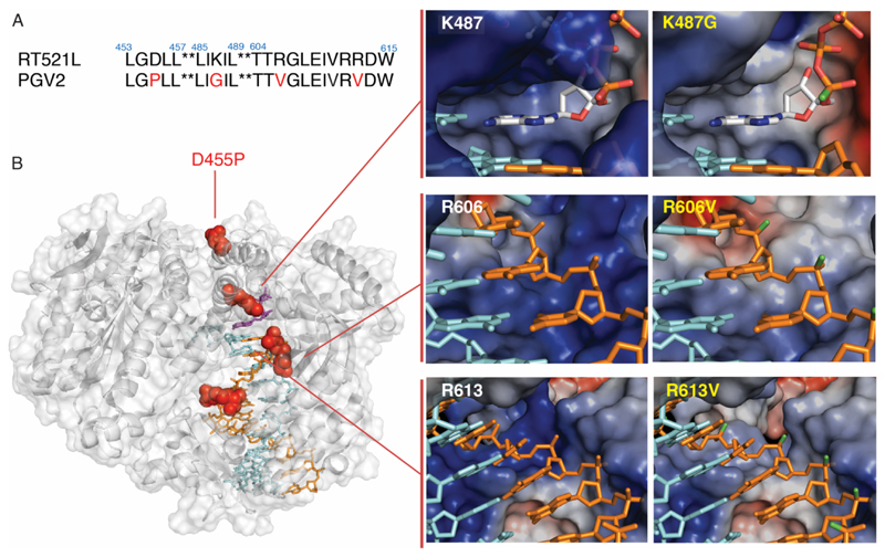
Figure 3. Polymerase mutations enabling phNA synthesis.
A) Sequence alignment of RT521L and phNA polymerase PGV2, mutated residues are highlighted in red. B) PGV2 mutations (red) mapped onto the structure of the ternary complex of the closely related KOD polymerase (PDB 5OMF) (left panel); surface structure and electrostatic surface potential changes upon mutation is shown for three mutations: K487G (top right), R606V (middle right) and R613V (bottom right panel) with nascent strand (orange), with or without Sp-P-alkyl substitution (green), and template strand (cyan).

