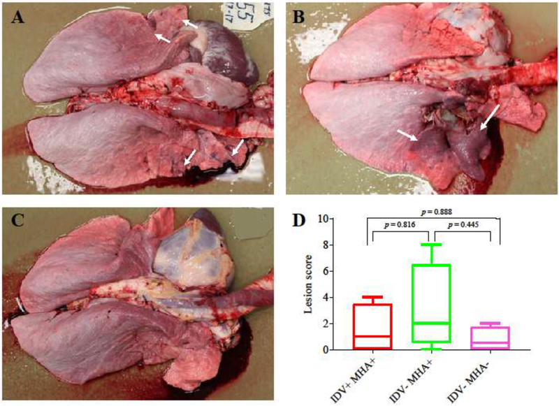
Fig. 2.
Gross pathology in the experimental and control cattle. Multifocal red areas (white arrows) were observed in the lungs of cattle from groups IDV+MHA+ (A) and IDV−MHA+ (B), but not in the lung of cattle from group IDV−MHA− (C); (D) Gross lung lesions were scored 0–3 based on the percentage of lung affected (0: no gross lesions; 1: <25% of lobe affected; 2: 25–75% of lobe affected; 3: >75% of lobe affected). All lung lobes were scored (right cranial, right cranial caudal, right middle, right caudal, left cranial, left cranial caudal, and left caudal) and the scores were added together to result in a gross lesion score (ranging from 0–8, with a possible maximum score of 21). The higher the gross pathology score, the more serious the pathogenesis. Box and whisker plots depicting gross pathology scores for IDV+MHA+, IDV−MHA+, and IDV−MHA− groups. A p-value of 0.05 was determined as statistically significant.

