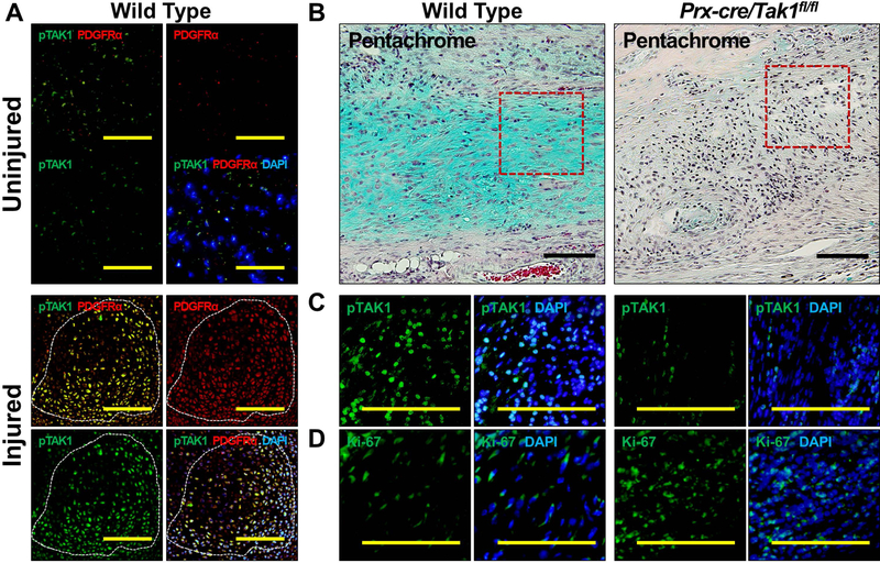
Figure 3. Genetic Loss of TAK1 signaling in mesenchymal cells increases cell proliferation and impairs chondrogenic differentiation to prevent trauma-induced HO.
(A) Co-expression of pTAK1 and PDGFRα+ in the injured and uninjured hindlimb 3 weeks after injury (20x magnification; Top Left Corner: pTAK1 – green overlay with PDGFRα – red; Bottom Left Corner: pTAK1 – green overlay with PDGFRα – red and DAPI - blue); (B) Pentachrome of injury site of Prx-cre/Tak1fx-frt/fx-frt and littermate control mice 3 weeks after injury (10x magnification; Alcian Blue represents cartilaginous tissue; red box shows areas of immunostaining); (C) Immunostaining for pTAK1 at the injury site of Prx-cre/Tak1fx-frt/fx-frt and littermate control mice 3 weeks after injury (10x magnification; Right Side: pTAK1 – green overlay with DAPI - blue); (D) Immunostaining for Ki67 at the injury site of Prx-cre/Tak1fx-frt/fx-frt and littermate control mice 3 weeks after injury (10x magnification; Right Side: Ki67 – green overlay with DAPI - blue). All scale bars = 200 μm.

