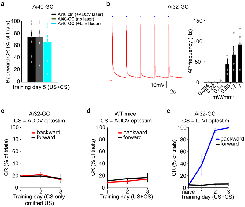
Extended Data Fig. 2. Optogenetic inactivation and stimulation of the ADCV and lobule VI in delay tactile startle conditioning.
(a) Day 5 backward conditioned responses of control mice subjected to delay tactile startle conditioning and optical stimulation in the ADCV as in Fig. 1i (Ai40 ctrl +ADCV laser, n=9 mice), Ai40-GC mice subjected to delay tactile startle conditioning in the absence of optical stimulation (Ai40-GC +no laser, n=6 mice), or Ai40-GC mice subjected to delay tactile startle conditioning and optogenetic silencing of lobule VI during the CS (Ai40-GC +L. VI laser, n=7 mice). (b) Acute sagittal cerebellar slices were prepared from mice expressing channelrhodopsin in granule neurons (Ai32-GC mice). Granule neuron action potentials were recorded in response to 100 ms optogenetic stimuli each composed of a 50 Hz train of 10 ms pulses (blue squares). A representative membrane potential trace for a granule neuron (left, n=2 neurons) and the relationship between the intensity of optostimulation of granule neurons and action potential firing (right, n=4 neurons). (c) Mice expressing channelrhodopsin in granule neurons (Ai32-GC mice) were subjected to optostimulation of the granule neuron pathway in the ADCV as the CS in the absence of the US (n=3 mice). (d) Control mice were subjected to optical stimulation in the ADCV as the CS together with the US (n=3 mice). (e) Mice expressing channelrhodopsin in granule neurons (Ai32-GC mice) were subjected to three days of delay tactile startle conditioning using optostimulation of lobule VI as the CS (backward conditioned responses in naïve vs day3, P=6.1×10⁻¹¹, two-tailed t-test, n=6 mice). In all panels, data show mean and error bars denote standard error.

