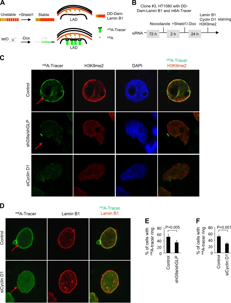
Fig. 8.
Cyclin D1 determines accumulation of G9a-mediated nuclear lamina interactions at the nuclear periphery. a Illustration of the inducible m6A-tracer/Dam-lamin B1 system in the HT1080 cell line stably expressing the Shield1-inducible Dam-Lamin B1 and the Tet-off m6A -tracer (known as the line clone 3). Inducible Dam-Lamin B1 expression was established by the fusion of a destabilization domain (DD), which causes Dam-Lamin B1 to be rapidly targeted for proteasomal degradation unless the protein is shielded by the synthetic small molecule Shield1.The induction of m6A-tracer is based on the Tet-Off system, where the removal of doxycycline (Dox) results in the activation of transcription. b Schematic representation of the experimental protocol. c Confocal microscopy images showing both G9a shRNA/GLP shRNA and cyclin D1 siRNA decreases H3K9me2 staining (red) in H1080 cells. d Representative confocal microscopy images showing cyclin D1 siRNA decreases accumulation of m6A-tracer signal (green) at the nuclear periphery after induction of Dam-Lamin B1. Lamin B1 staining is shown in red. e Quantitative analysis of m6A-tracer incorporation at the nuclear periphery after induction of Dam-Lamin B1 expression shown as % of cells incorporating tracer for n > 200 separate cells for shG9a/shGLP treatment and its vector control. Data are percentage ± 95% confidence interval (CI). f Quantitative analysis of m6A-tracer incorporation at the nuclear periphery shown as % of cells incorporating tracer for n > 400 separate cells cyclin D1 siRNA treatment as well as its control. Data are percentage ± 95% CI

