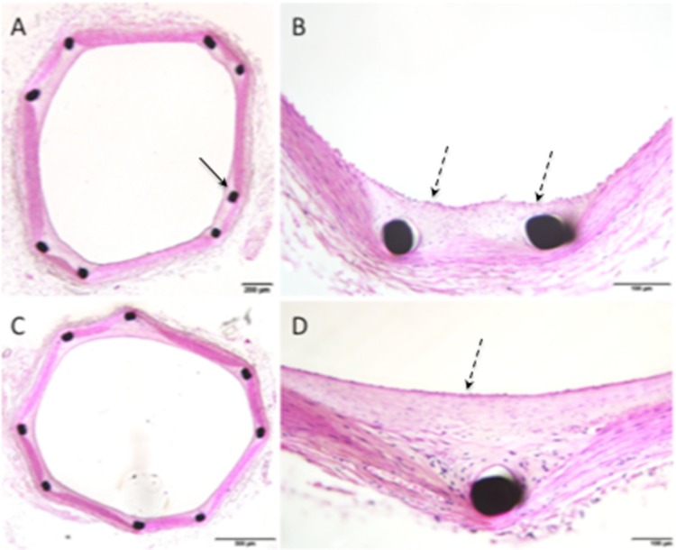
Fig. 3.
Histological imaging of animal 11, performed at 180 days post-implantation, showed SMC rich neointima (dashed black arrows) completely covering the struts (solid black arrow) of the pCONUS (A, B) and pCONUS HPC (C, D) with no evidence of chronic inflammation. There was no evidence of adventitial inflammation

