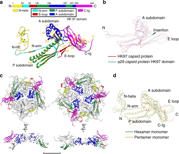
Fig. 2.
Structure of the mature capsid. a Structure of the mature virion major capsid protein gp8. Top: a schematic diagram showing the polypeptide chain of a gp8 monomer; bottom: a ribbon diagram of a gp8 monomer structure in a pentameric capsomer. The structural domains of gp8 are shown in yellow (N-helix), cyan (HK97-N arm), red (HK97-E loop), green (HK97-P), blue (HK97-A), and magenta (C-Ig). b Structural superposition of the ϕ29 HK97 domain (blue) and the HK97 capsid protein (red). c Top and side view ribbon diagrams of a pentameric capsomer and a hexameric capsomer. The structural domains of two gp8 monomers in the pentameric capsomer and three gp8 monomers in the hexameric capsomer are colored in the same manner as in a. The scale bar represents 5 nm. d Structural superposition of a pentameric capsomer monomer (orange) and a hexameric capsomer monomer (green)

