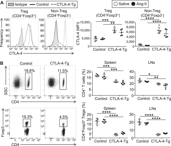
Figure 3.
Overexpression of cytotoxic T lymphocyte-associated antigen-4 (CTLA-4) decreases the frequency of CD4+ T cells and CD4+ forkhead box P3 (Foxp3)+ regulatory T cells (Tregs) in angiotensin II-treated or saline-treated mice. Seven days after the pump implantation, lymphoid cells from spleen and lymph nodes (LNs) were prepared. Apolipoprotein E-deficient (Apoe−/−) mice infused with angiotensin II or saline served as controls. (A) Representative results of intracellular CTLA-4 expression in splenic CD4+Foxp3+ Tregs and CD4+Foxp3− T cells of angiotensin II-infused Apoe−/− and CTLA-4-Tg/Apoe−/− mice assessed by flow cytometry. The expression levels of intracellular CTLA-4 were examined. The data are shown as mean fluorescence intensity (MFI). n = 5 per group. (B) Representative results of Foxp3 and CD4 expression in the spleen of angiotensin II-infused Apoe−/− and CTLA-4-Tg/Apoe−/− mice assessed by flow cytometry. The graphs represent the percentage of CD4+ T cells or CD4+Foxp3+ Tregs within spleen and LN cells or spleen and LN CD4+ population, respectively. n = 5 mice per group. Data points represent individual animals. Horizontal bars represent means. Error bars indicate s.d. *P < 0.05; **P < 0.01; ***P < 0.001; ****P < 0.0001; one-way ANOVA followed by Tukey’s post hoc test. Ang-II, angiotensin II; SSC, side scatter.

