Fig. 5.

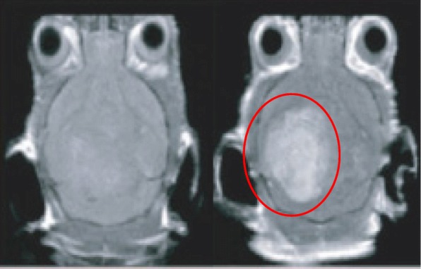
An MRI contrast image of a rat cerebral cortex pre- (left) and post-treatment (right). The area containing the AuNPs is ringed in red

An MRI contrast image of a rat cerebral cortex pre- (left) and post-treatment (right). The area containing the AuNPs is ringed in red