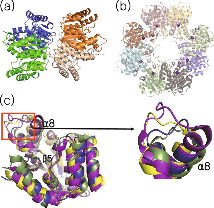
Figure 2.
Overall structures of Rv0187. Ribbon representation of crystal structures of (a) ligand-free and (b) cofactor-bound forms of Rv0187, where SAH and the strontium ion are shown as white gray sticks and a purple sphere, respectively. Shown is the composition of the asymmetric unit, with four and eight copies of monomer in (a) and (b), respectively. (c) Conformational comparison of Rv0187 with its three closest structural homologs. Each monomer is superimposed, and ligand-free Rv0187 is presented in blue, the putative OMT of C. glutamicum (PDB code 3DR5) is yellow, S. achromogenes TomG (5N5D) is green, and S. bicolor CCoAOMT (5KVA) is purple. Inset is a magnified view of the region where the largest conformational diversity occurs.

