
Figure 1.
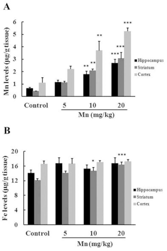
Effects of Mn exposure on metal accumulation in the hippocampus, striatum and cerebral cortex of immature rats. The panels show the accumulation of Mn (A) and Fe (B). Rat pups were treated with saline (control; NaCl 0.9%) or MnCl2 at doses of 5, 10 or 20 mg/kg for twenty days (PN8–27) and the brain metal concentrations were analyzed on PN29. Results represent mean ± S.E.M and are expressed in ĝ metal/g tissue. Statistical analysis was performed by ANOVA followed by Duncan’s test. n = 4; * p < 0.05, ** p < 0.01, *** p < 0.001 compared to control.