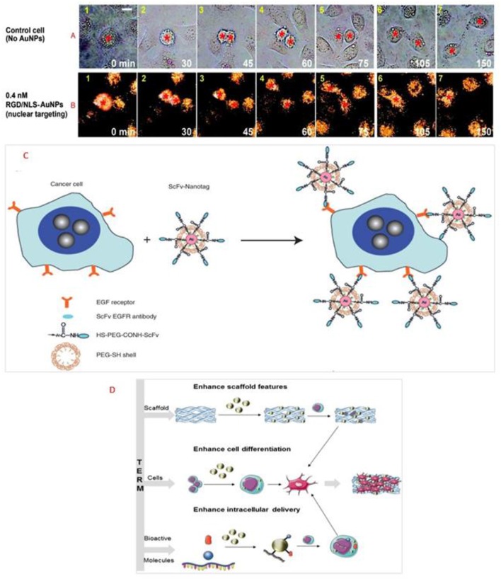
Figure 2.
Real-time images of cancer cell division under the following conditions: (A) with No AuNPs and (B) in the presence of 0.4 nM nuclear-targeting gold nanoparticles (RGD/NLS-AuNPs). Red stars indicate the nuclei. Scale bar: 10 μm. Reprinted from Kang et al. (2010) with permission from American Chemical Society. (C) Preparation of targeted surface-enhanced Raman scattering (SERS) NPs by using a mixture of SH-PEG and a hetero-functional PEG (SH-PEG-COOH). Covalent conjugation of an EGFR-antibody fragment occurs at the exposed terminal of the hetero-functional PEG. Reprinted from Qian et al. (2008) with permission from John Wiley and Sons. (D) Scheme representing the use of AuNPs in tissue engineering and regenerative medicine. Reprinted from Vial et al. (2017) with permission from Elsevier.

