Figure 5.

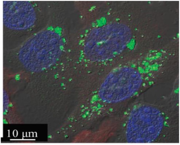
A merged fluorescence confocal image of FMN–MTNs (green dots) in BT-20 cancer cells where the nuclei were stained with DAPI (blue spots). Reprinted from Wu et al. (2011) with permission from The Royal Society of Chemistry.

A merged fluorescence confocal image of FMN–MTNs (green dots) in BT-20 cancer cells where the nuclei were stained with DAPI (blue spots). Reprinted from Wu et al. (2011) with permission from The Royal Society of Chemistry.