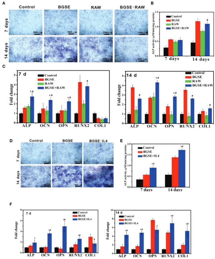
Figure 9.
(A-C) Osteogenic differentiation of 3T3 cells under inflammatory state. (A) ALP staining and (B) ALP quantitative assay of 3T3 cells after cultured for 7 and 14 days. (C) mRNA expression of osteogenesis-related genes (ALP, OCN, OPN, COL1, RUNX2) in 3T3 cells cultured for 7 and 14 days. *p < 0.05 vs. BGSE (bioactive glass scaffolds extracts) group; #p < 0.05 vs. RAW (macrophage-like cell line) group. (D–F) Osteogenic differentiation of 3T3 cells in anti-inflammatory state. (D) ALP staining and (E) ALP quantitative assay of 3T3 cells after being treated with various samples for 7 and 14 days. (F) mRNA expression of osteogenesis-related genes (ALP, OCN, OPN, COL1, RUNX2) of 3T3 cells incubated with various groups for 7 and 14 days. *p < 0.05 vs. blank control; #p < 0.05 vs. BGSE group. Reproduced from Zhao et al. (2018) with permission from John Wiley and Sons.

