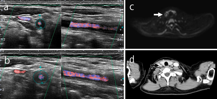
Figure 1.
Ultrasonography showed a hypoechoic halo surrounding the right common carotid artery (a) on the day of admission. However, no significant changes were observed compared to four weeks prior (b). DWIBS (c) and contrast CT (d) showing inflammation on the same artery wall for which ultrasonography showed a halo on the day of admission. DWIBS: diffusion-weighted whole-body imaging with background body signal suppression

