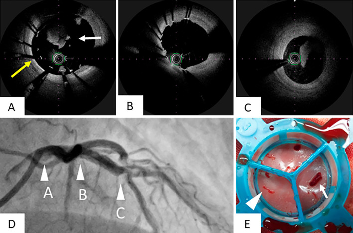
Figure 3.
Findings of CAG and OFDI 2 days later after primary PCI. We performed CAG and OFDI two days after primary PCI. In OFDI, we noted remnant thrombi (A, white arrow) and incomplete stent apposition in the LM (A, yellow arrow) and no thrombi and complete apposition in the LAD (B). Intimal dissection and thrombi were observed near the stent distal edge (C). The arrowheads of A, B, and C in Fig. D showed points of OFDI views of Fig. A, B, and C. The thrombus was retrieved using an aspiration catheter from the coronary artery (E, arrowhead) and guiding catheter (E, small arrow).

