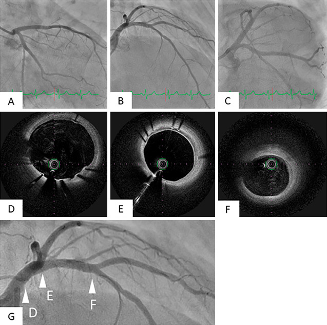
Figure 4.
Findings of follow-up CAG and OFDI nine months later. Follow-up CAG nine months later revealed no in-stent restenosis. In addition, the moderate stenosis in the mid LAD had improved (A, B, C). OFDI showed that the stent was completely covered with neointima over the full length with no thrombi from the LM to the LAD. (D, E, F). The arrowheads of D, E, and F in Fig. G showed points of OFDI views of Fig. D, E, and F.

