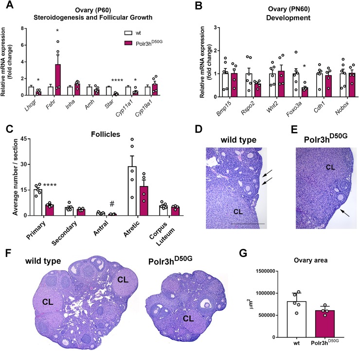
Figure 7.
Polr3h D50G mice show changes in ovarian transcript levels and decreased number of primary follicles. (A) Relative expression of genes associated with steroidogenesis and follicular growth. Note decreased levels of Lhcgr, Star, and Cyp11A1 and increased Fshr in Polr3hD50G female mice (df = 9; t > 2.4 for differences). (B) Relative expression of genes associated with ovary development. Note decreased levels of Foxo3a in Polr3hD50G female mice (df = 9, t > 2.38 for differences). (C) Average number of follicles per section in different stages of development. Note decreased number of primary follicles (df = 7, t = 5.9). Differences were determined by multiple t test (P < 0.05). Discovery was determined via the two-stage linear step-up procedure of Benjamini, Krieger, and Yekutieli. (D–F) Bright field images showing sections of ovaries. Arrows point to primary follicles (D, E). (G) Graph showing average sectional ovary area for both genotypes. Two-tailed Student t test was used (P = 0.08). *P < 0.05; ****P < 0.001, #P < 0.05 but q > 0.1. Amh, anti-Mullerian hormone; Bmp15, bone morphogenetic protein 15; Cdh1, cadherin 1; CL, corpus luteum; Cyp11a1, cytochrome P450 family 11 subfamily A member 1; Cyp19a1, cytochrome P450 family 19 subfamily A member 1 (aromatase); Foxo3, forkhead box O3; Fshr, follicle stimulating hormone receptor; Inha, inhibin a; Lhcgr, luteinizing hormone/chorionic gonadotropin receptor; Nobox, NOBOX oogenesis homeobox; Rspo2, R-spondin 2; Star, steroidogenic acute regulatory protein; Wnt2, Wnt family member 2.

