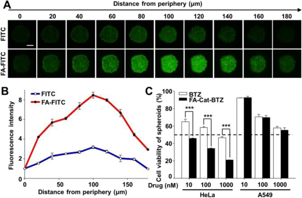
Fig. 7.

FA-mediated penetration and cytotoxicity in 3D cancer cell spheroids. (A) Spheroid penetration by FITC and FA-FITC, determined by confocal microscopy. Incubation time: 1.5 h. (B) Quantitation of the fluorescence intensity of the images by the ImageJ software. (C) Cell viability of the BTZ conjugate in the spheroids, determined by the CellTiter-Blue® Cell Viability Assay. Incubation time: 48 h. Scale bar: 200 μm.
