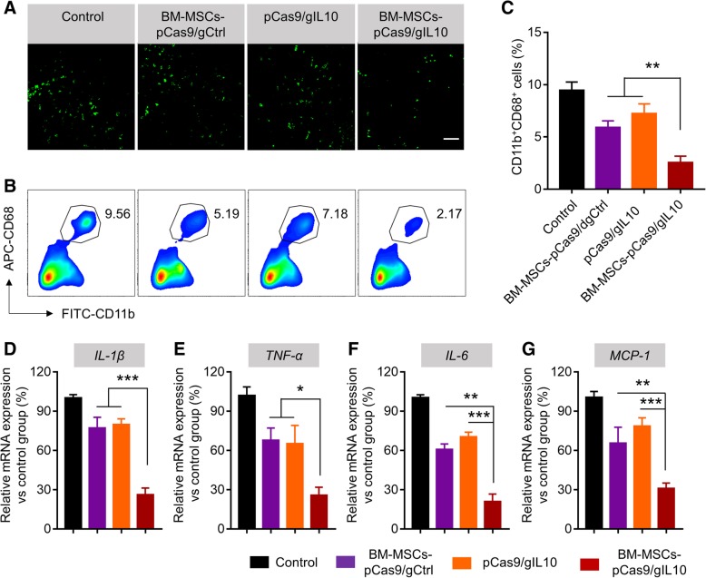
Fig. 3.
Transplantation of BM-MSCs-pCas9/gIL10 suppresses infiltration of inflammatory cells and expression of proinflammatory cytokines in the myocardium. a Representative fluorescent micrographs show the presence of inflammatory cells (CD68-positive, green fluorescence) peri-infarct area in the control MI heart or hearts treated with BM-MSCs pCas9/gCtrl, pCas9/gIL10 or BM-MSCs pCas9/gIL10. The examination was performed 1-week post transplantation. Scale bar was 20 μm. b Flow cytometry analysis of infiltration of CD11b+CD68+ cells peri-infarct area in the heart. Tissues from control MI heart or hearts treated with BM-MSCs-pCas9/gCtrl, pCas9/gIL10 or BM-MSCs-pCas9/gIL10 were digested into single cells stained with FITC labeled CD11b and APC labeled CD68 antibodies, and then examined by flow cytometry. c Bar graph shows quantitative analysis of infiltrating CD68-positive cells. Data represent means ± SD. **p < 0.005, n = 8. d-g Quantitative analysis of mRNA expression of proinflammatory cytokines and chemokines (IL-1β, TNF-α, IL-6 and MCP-1) in the border zone of LV infarct of LV infarct at 1-week post-MI. mRNA expression normalized to GAPDH expression. Data represent means ± SD. *p < 0.05, **p < 0.005, ***p < 0.001, n = 8

