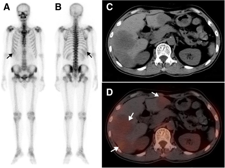
Fig. 1.

A 62-year-old man with rectal cancer after surgery and chemotherapy for 3 years, who was referred for suspected metastasis. The whole-body scan (a, anterior; b, posterior) demonstrated 99mTc-MDP uptake in right upper abdomen (black arrow). Axial CT (c), and SPECT/CT images (d) showed increasing 99mTc-MDP uptake corresponding to multiple liver metastases without calcification (white arrow)
