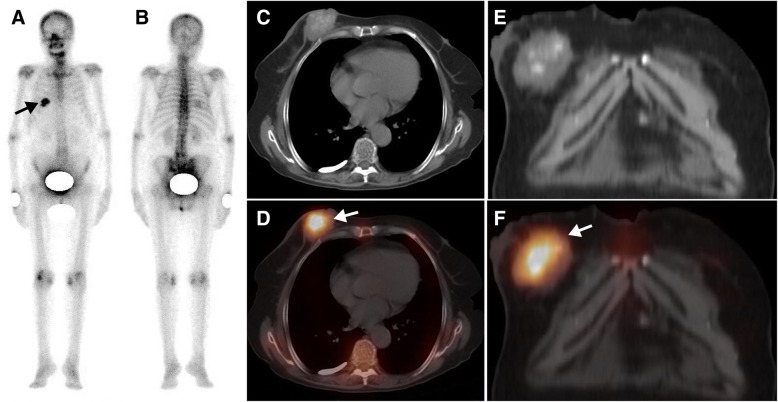
Fig. 2.
A 76-year-old woman with breast cancer was referred for metastatic work-up before surgery. The whole-body scan (a, anterior; b, posterior) revealed a focal area of high 99mTc-MDP activity in the right 5-6th posterior rib region (black arrow). Axial CT (c), and coronal CT (e) and SPECT/ CT images (d, f) showed that elevated 99mTc-MDP activity was located in a breast tumour with calcification (white arrow)

