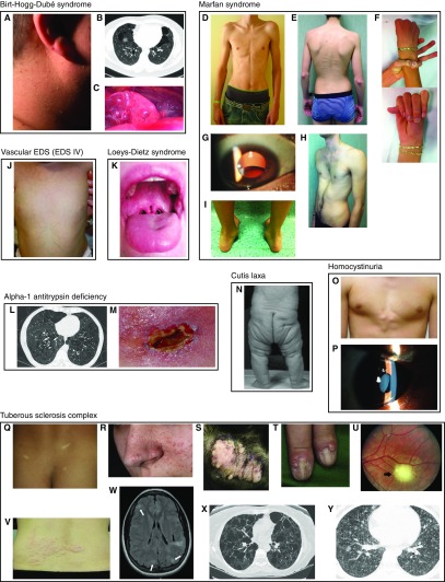
Figure 2.
Physical examination findings of pneumothorax-associated syndromes. (A–C) Birt-Hogg-Dubé syndrome. (A) Fibrofolliculomas of the neck (159). (B) Lung cysts and bullae; extra-apical location is characteristic (159). (C) Pleural blebs on the surface of the left lower lobe; these can be missed on computed tomography but seen during thoracoscopy/video-assisted thorascopic surgery (159). (D–J) Marfan syndrome. (D) Marfanoid body habitus (90). (E) Scoliosis, striae, reduced elbow extension (160). (F) Positive thumb and wrist signs indicating arachnodactyly (160). (G) Lens dislocation (161). (H) Pectus excavatum (160). (I) Hindfoot deformity (160). (J) Vascular (type IV) Ehlers-Danlos syndrome: translucent skin on the torso of an infant (162). (K) Loeys-Dietz syndrome: bifid uvula (163) (L and M) Alpha-1 antitrypsin deficiency. (L) Panlobular emphysema (case courtesy of Dr. Jeremy Jones, Radiopaedia.org, rID:13441). (M) Panniculitis (164). (N) Cutis laxa: autosomal recessive cutis laxa IIa (165). (O and P) Homocystinuria. (O) Chest wall deformity (166). (P) Lens dislocation (167). (Q–Y) Tuberous sclerosis complex. (Q) Hypopigmented macules (ash leaf spots) (168). (R) Angiofibromas (168). (S) Fibrous plaques (168). (T) Ungual fibromas (168). (U) Retinal hamartoma (168). (V) Shagreen patch (168). (W) Cortical dysplasia (tubers, white arrows; radial migration lines, black arrow) (168). (X) Lymphangioleiomyomatosis (168). (Y) Multifocal micronodular pneumocyte hyperplasia (169). Images are reproduced by permission from the sources cited above.

