. 2018 Mar 30;35(1):59–70. doi: 10.5511/plantbiotechnology.18.0202a

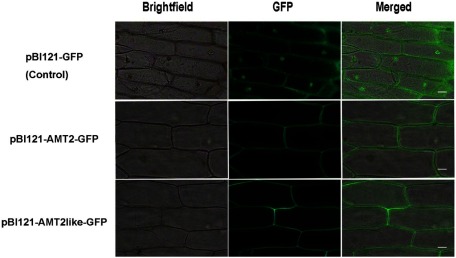
Figure 3. Subcellular localization of pBI121-GFP, pBI121-BcAMT2-GFP, and pBI121-BcAMT2like-GFP fusion proteins in onion epidermal cells. Bright field: image obtained by bright field microscopy; GFP: green fluorescence derived from GFP imaged by fluorescence microscopy; Merged: overlay of the two images. Scale bar: 50 µm.

Figure 3. Subcellular localization of pBI121-GFP, pBI121-BcAMT2-GFP, and pBI121-BcAMT2like-GFP fusion proteins in onion epidermal cells. Bright field: image obtained by bright field microscopy; GFP: green fluorescence derived from GFP imaged by fluorescence microscopy; Merged: overlay of the two images. Scale bar: 50 µm.