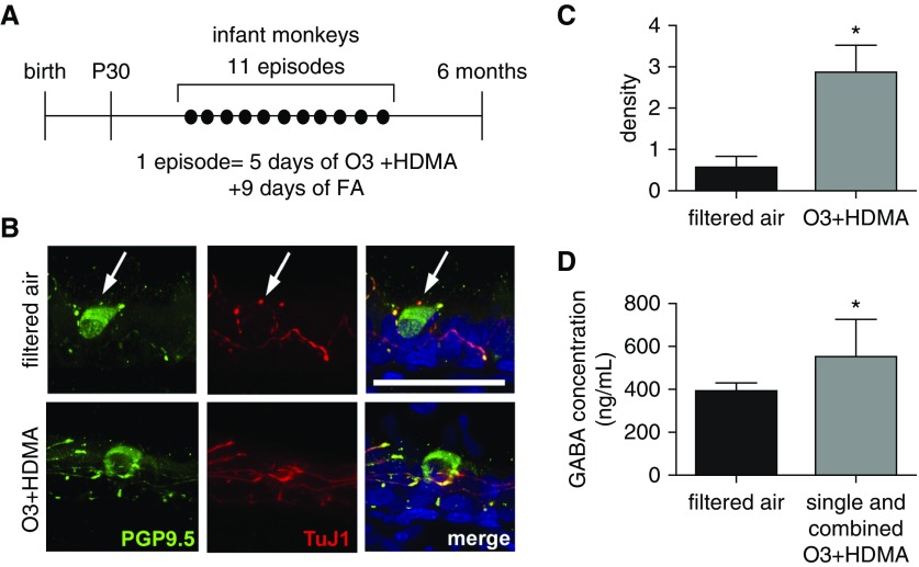
Figure 2.
PNEC hyperinnervation and GABA hypersecretion in a nonhuman primate model of early-life exposure. (A) Experimental scheme of rhesus monkey ozone (O3) and house dust mite allergen (HDMA) exposure during the first 6 months after birth. Control animals were exposed to filtered air (FA). Lungs and serum samples were harvested at 6 months of age. (B) Representative Z-stack confocal images of PNEC innervation in 6-month-old rhesus monkeys with and without O3 + HDMA exposure. PNEC innervation was assessed by double staining of proximal lung sections (25 μm in thickness) for PGP9.5 and neuron-specific β-tubulin III using a TuJ1 antibody. Arrows mark PNECs. Scale bar: 50 μm. (C) Quantification of PNEC innervation in infant rhesus monkeys with and without O3 and HDMA exposure. The density of nerves surrounding PNECs was calculated by normalizing TuJ1-immunoreactive area to the number of PNECs. At least five single PNECs and PNEC clusters from three lungs in each group were measured. (D) Serum concentrations of GABA in control animals (n = 7) and rhesus monkeys (n = 10) that were exposed to O3, HDMA, and combined O3 + HDMA. Data presented in C and D represent mean ± SEM. *P < 0.05.

