Figure 1.
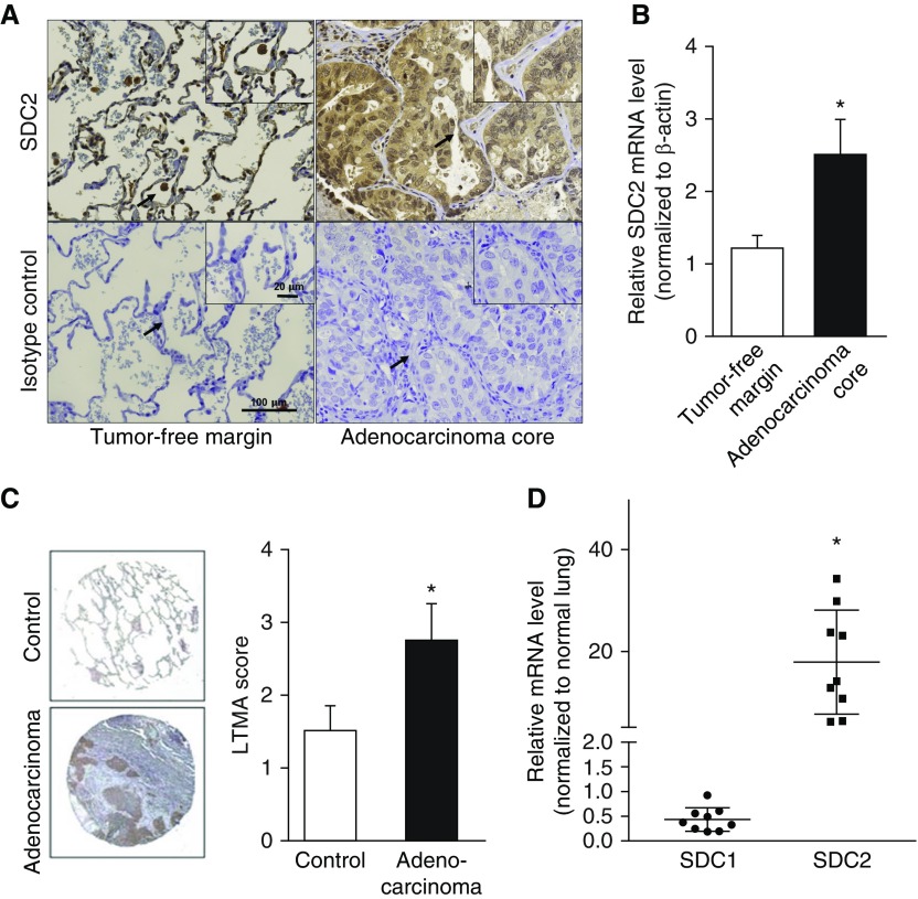
Syndecan (SDC)-2 is overexpressed in human lung adenocarcinoma. (A) SDC2 expression in lung adenocarcinoma tumor core and tumor-free margins was assessed by immunohistochemistry. SDC2 expression was detected in tumor-affected epithelium (upper right panel) and tissue-associated macrophages in tumor-free margins (upper left panel). Representative images are shown. The arrows indicate the area of magnification. (B) SDC2 mRNA levels were elevated in tumor core samples compared with tumor-free margin samples (n = 16; *P < 0.05). (C) Anti–human SDC2 antibody was applied to a Lung Cancer Tissue MicroArray (LTMA) in control lung tissue (n = 8) and lung adenocarcinoma (n = 24). Staining intensity was scored from 1 to 4 (lowest to highest, respectively), showing increased staining in adenocarcinoma tissue compared with controls (*P < 0.05). Representative images are shown. (D) SDC1 and SDC2 mRNA levels were measured in human lungs with adenocarcinoma and control lungs without cancer (n = 9). Data represent fold increase of SDC2 expression relative to normal lung tissue. SDC2 levels, but not SDC1 levels, were increased 5- to 35-fold compared with controls (*P < 0.05). Scale bars: 20 μm and 100 μm.

