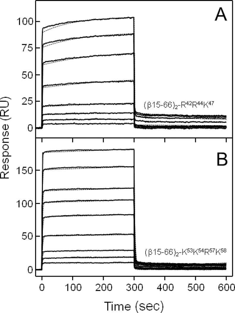
Figure 8.

Analysis of interaction of the (β15–66)2 Lys-Arg-mutants with the fibrin-binding VLDLR(1–8) fragment by SPR. The (β15–66)2-R42R44K47 mutant at increasing concentrations, 0.1, 0.25, 0.5, 1.0, 2.5, 5, 7.5, and 10 μM (A), or the (β15–66)2-K53K54R57K58 mutant at 0.25, 0.5, 1.0, 2.5, 5, 7.5, 10, 15, and 20 μM (B) was added to the immobilized VLDLR(1–8) fragment and its association/dissociation was monitored in real time while registering the resonance signal (response) using BIAcore biosensor. The dotted curves in both panels, which essentially coincide with the experimental curves, represent the best fit of the binding data using global fitting analysis (see Experimental Procedures). The Kd values determined from SPR binding data are presented in Table 3.
