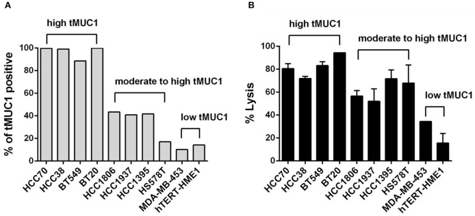
Figure 2.
The MUC28z CAR T cells lyse TNBC tumor cells in vitro in an antigen-dependent manner. (A) Percentage of cells expressing tMUC1, determined by TAB004-APC/Cy5.5 staining and flow cytometry. A panel of nine TNBC cell lines and one “normal” mammary epithelial cell line hTERT-HME1 is shown. (B) Percentage of TNBC tumor cell lysis by MUC28z CAR T cells. Cells were co-cultured at E:T ratio of 5:1 for 3 days. The lysis of tumor cells was determined by MTT assay. Data are presented as the mean ± SEM. The relationship between tMUC1 positivity in TNBCs and tumor lysis by MUC28z CAR T cells was analyzed by a non-parametric Spearman correlation, with r = 0.8909 which was highly significant (P = 0.0011) and indicated a positive association between tMUC1 level and tumor lysis.

