Figure 4.
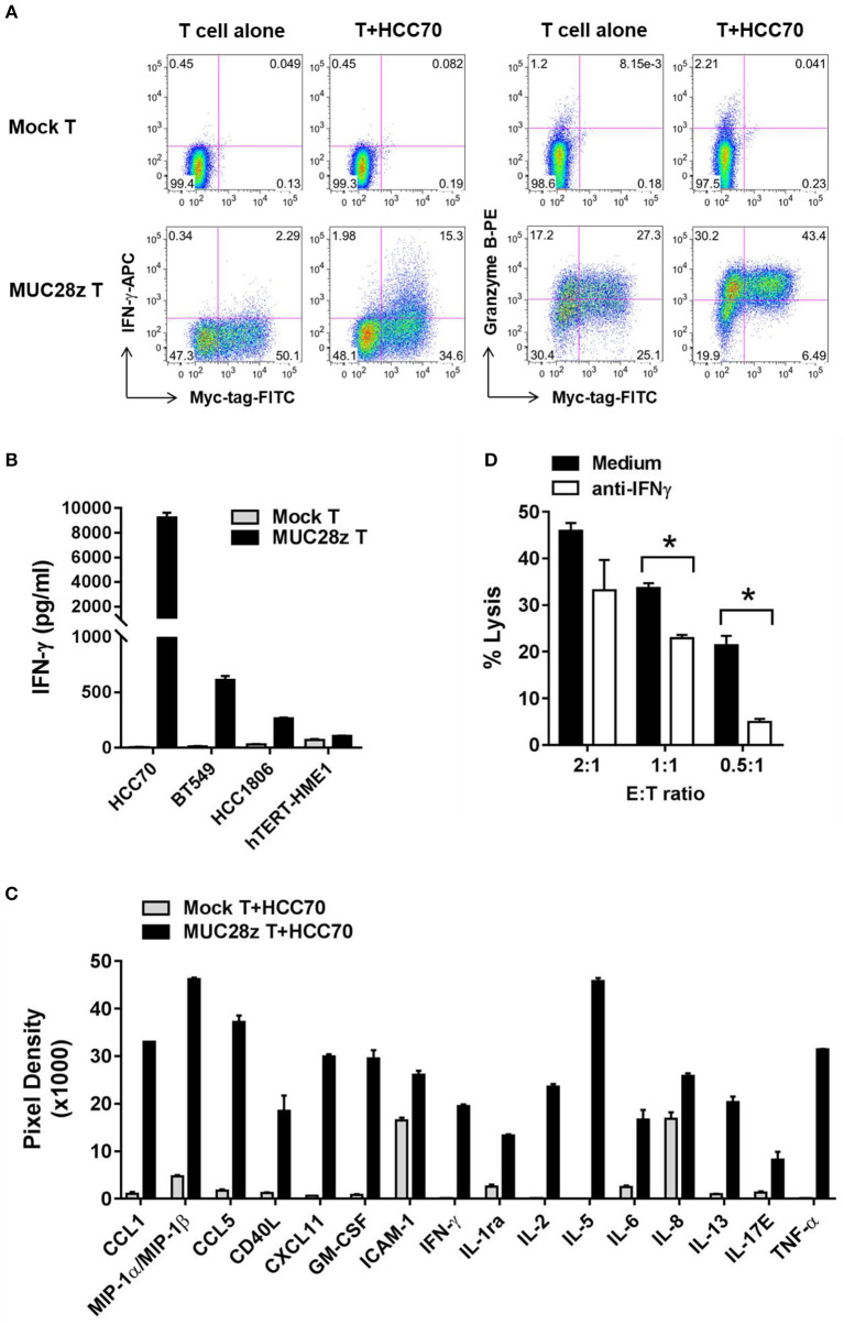
MUC28z CAR T cells release antigen-specific cytokines when co-culture with tMUC1high TNBCs. (A) CD8+ MUC28z CAR T cells produced IFN-γ and increased Granzyme B in response to tMUC1high HCC70 cells in vitro. MUC28z CAR T cells were co-cultured with HCC70 tumor cells for 24 h, and then cells were stained intracellularly for IFN-γ and Granzyme B in addition to Myc-tag. Cells were gated on CD8+ T cells, followed by flow cytometry analysis. (B) MUC28z CAR T cells produced IFN-γ in response to tMUC1-expressing TNBC in vitro. T cells were co-cultured with the selected tumor cell lines (E:T = 2:1) for 24 h, and then the culture supernatants were assayed for IFN-γ by ELISA. Data are presented as mean ± SD of replicates. The baseline IFN-γ releases in the absence of TNBC stimulation are: mock T = 2.6 ± 0.9 pg/ml; MUC28z CAR T = 18.7 ± 0.1 pg/ml. (C) MUC28z CAR T cells released a large panel of cytokines after tMUC1-specific cell activation. T cells were co-cultured with HCC70 cells (E:T = 2:1) for 24 h, and then the culture supernatants were assayed for cytokine concentration by human cytokine array. Data are presented as mean ± SD of replicates. (D) The lysis of HCC70 cells by MUC28z CAR T cells was partially reversed by IFN-γ neutralization. HCC70 cells were co-cultured with MUC28z CAR T cells for 24 h in the absence or presence of IFN-γ neutralizing antibody. The lysis of HCC70 cells was determined by MTT assay. Data are presented as the mean ± SEM. *p < 0.05 (student t-test).

