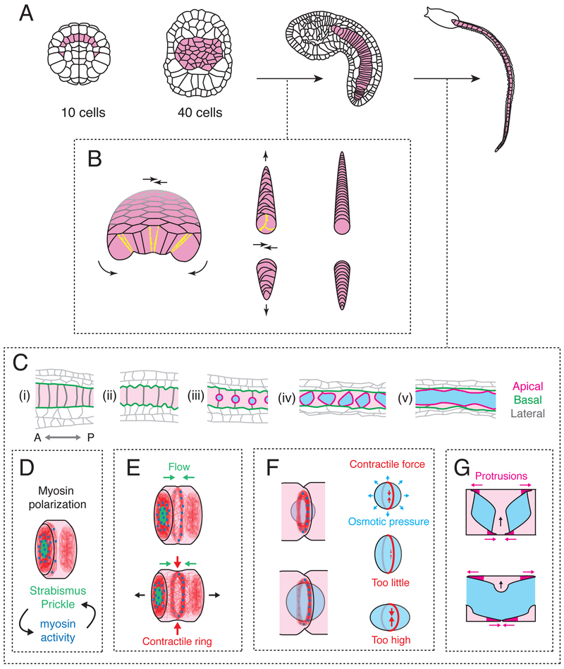
Figure 2. Cytomechanical basis for notochord elongation and tubulogenesis.
(A) The notochord transforms from a set of ten precursor cells into a monolayer plate, then a cylindrical rod, then an elongated tube. (B) Transformation of the flat notochord plate into a cylindrical rod of coin-shaped cells is driven by basolateral crawling (yellow protrusions), which is polarized in the apico-basal plane to drive bending/invagination (curved black arrows) and planar polarized to drive convergent extension (straight black arrows). Modified from [5]). (C) Stages of notochord elongation and tubulogenesis. Schematics show a sagittal section along the notochord midline. Anterior is to the left. Cyan regions indicate lumen; apical, basal and lateral domains are shown in magenta, green and grey respectively. (D) Before elongation, polarized accumulation of Myosin II (blue) and PCP proteins (green) on anterior lateral cell contacts is governed by their reciprocal interactions. A basal actomyosin ring (red filaments and blue Myosin II) forms at the anterior edge of each cell. (E) Anterior enrichment of Myosin II and PCP proteins persists, and a self-centering cortical flow positions the basal actomyosin ring at the cell equator, where it contracts to drive cell elongation. (F) A peri-apical actomyosin contractile ring at cell contacts counteracts osmotic forces to control lumen expansion. (G) Bidirectional polarized basal crawling of notochord cells drives fusion of individual lumens into a central core.

