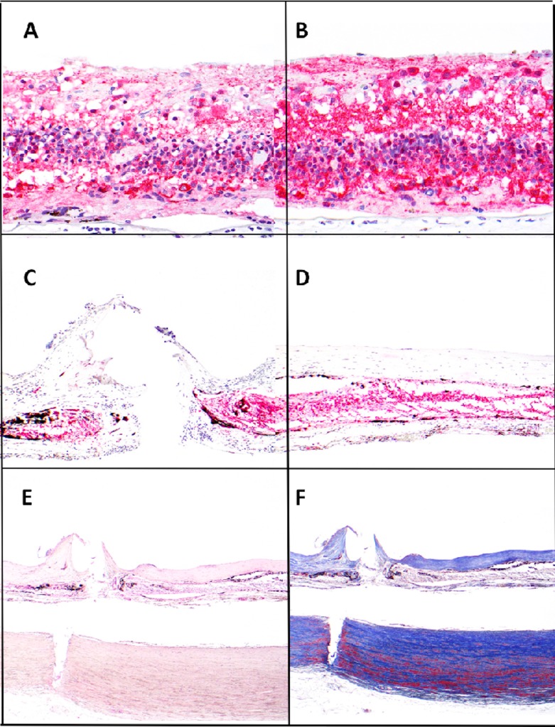
Figure 5.
Sections of retina from implanted and fellow eyes. Consistent highly remodeled retina was found across all sections. Retinal neuron counts were calculated from NSE staining (A–D) and tack penetration shown by H&E and Masson Trichrome staining (E, F). The total neuron counts of the macular area showed no significant difference between both eyes. ([A] implant eye, [B] fellow eye, NSE, ×400). Significant loss of neurons was only revealed at and near the tack with fibrosis formation ([C] tack site, [D] adjacent areas with fibrotic membrane, NSE, ×100). Deposition of dense collagen in the fibrotic membrane was readily identified with H&E and Masson Trichrome stain. The tack sites showed an all layer penetrated wound from the retina through the choroid, and to the outer lamina of the sclera ([E] H&E, ×40, [F] Masson Trichrome, ×40).

