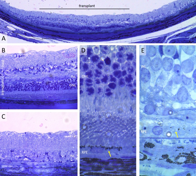
Figure 2.
Histology of transplanted retina 2 weeks postsurgery. (A) Wide view of the transplanted retina (LE transplanted into RCS rat) showing the 1-mm transplant (black line) and the control area outside it. (B, C) Higher magnification of the same sample showing excellent preservation of the transplanted photoreceptors (B), compared with the area outside the transplant (C). The inner retina of the transplant is significantly thinned, with no distinct GCL. (D, E) The close up illustrates pigmented RPE monolayer under the transplant (yellow arrow [D]), as opposed to nonpigmented RPE in a control area (E).

