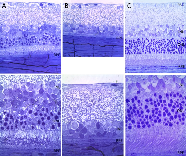
Figure 4.
Histology of a transplanted retina at 6 months postoperative, compared with control and with wild-type retina. (A) The transplanted area has nearly normal anatomy, with a single INL and transplanted photoreceptors merging with the host retina (from a LE into S334ter rat). (B) The nontransplanted area completely lacking photoreceptors. (C) A healthy LE rat retina is shown here for comparison. Top row is imaged via ×40 objective and bottom row via ×100.

