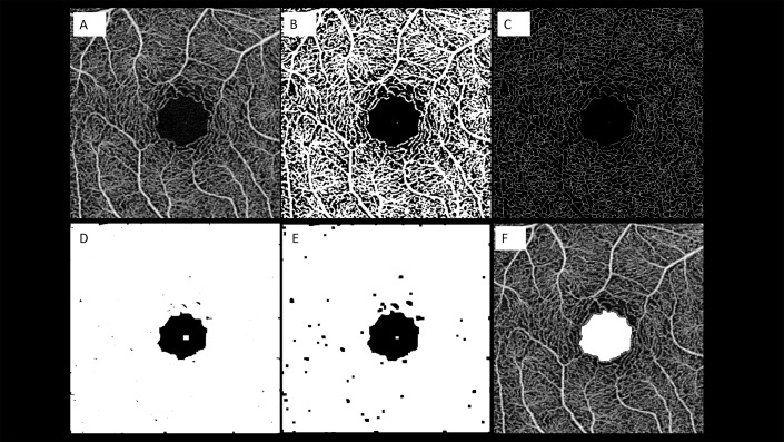
Figure 3.
Representation of the algorithm used to process the images. The images of the superficial capillary plexus (SCP) were first imported in ImageJ software. After running the erode command on the en face image, which we acquired in 800 × 800 pixels (A), we performed binarization (B) and skeletonization (C). Subsequently, we repeated the dilate command for connecting capillary ring disruption (D) and repeated the erode command for narrowing vessels (E). Finally, we returned the image size to normal and extracted the FAZ line (F).

