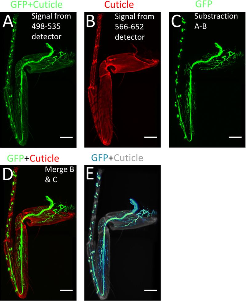
Figure 4: Processing of images using ImageJ/FIJI.

(A) Max projection of the confocal stacks obtained from 498–535 nm detector. (B) Max projection of the confocal stack obtained from 566–652 nm detector. (C) Image with only GFP signal obtained by subtracting (A) from (B). (D) Merged images of (B) and (C). (E) 3-D reconstruction of (C). Scale bar = 100 μm.
