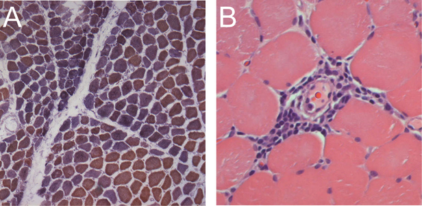
Figure 1.
Typical muscle biopsy from an anti-Tif1γ positive DM patient. (A) This low power view of a frozen section stained with COX (brown) and SDH (blue) reveals both normal fibers (brown) and numerous COX-deficient fibers (purple/blue) indicating mitochondrial dysfunction; several fascicles include examples of perifascicular atrophy. (B) This high power view of a paraffin section from the same patient stained with H&E shows a striking example of perivascular inflammation.

