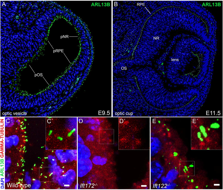
Fig. 1.
Optic progenitor cells are ciliated and require IFT122 and IFT172 for proper cilia assembly. (A-B) Sections through a wild-type E9.5 optic vesicle (A) and a wild-type E11.5 optic cup (B). Cilia are visualized with an antibody against ARL13B (green) and DNA counterstained with DAPI (blue). Abbreviations for optic progenitor territories: pOS, presumptive optic stalk; pRPE, presumptive retinal pigment epithelium; pNR, presumptive neural retina; OS, optic stalk; RPE retinal pigment epithelium; NR, neural retina. (C-E) Maximum intensity projections of confocal z-stacks of wild-type (C), Ift172 mutant (D) and Ift122 mutant (E) eyes in the presumptive optic stalk at E10.5 stained with antibodies against ARL13B (green) and γ -TUBULIN (red); DNA counterstained with DAPI (blue). Scale bars are 2 μm. Insets (C’-E’) are magnifications of the area marked by the dotted box in the corresponding image (C-E).

